 Hablamos Español
Mixed Oligodendroglioma-Astrocytoma
This unusual brain tumor is a mixture of two different lines of tumor cells originating from two different support cells that surround the neurons: oligodendroglial cells and astrocytes. The oligodendroglial cells produce the myelin sheaths that insulate the nerve fibers in the white matter, and astrocytes are support cells that play a nutritional role to the neurons. The tumors exhibit behavior that parallels the astrocytic component. The pathologic grade of the astrocytic component determines the overall behavior of the whole tumor.
The example below shows a 40 yr old patient who developed headaches and seizures. The original imaging showed a large tumor measuring 7.2 cm in diameter located in the right frontal area which exerted marked compression of the adjacent brain tissue.
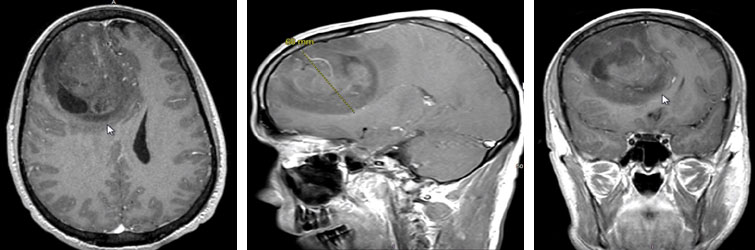
The arrows point to the interface of the tumor with adjacent brain on 1. axial, 2. sagittal, and 3. coronal plane MRI slices.
Microsurgical resection was performed.

In the image above, the brain is seen to be full and under pressure. The normal sulcal-gyral pattern of folds on the brain surface is obliterated due to the pressure of the tumor expanding the brain tissue.
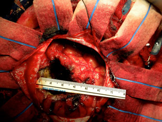
After microsurgical resection of the tumor, the brain is now visibly decompressed. It is helpful to measure the size of the cavity after tumor removal to help correlate the volume resected with the preoperative measurements of the tumor on imaging. This helps to minimize the possibility of inadvertently leaving some tumor behind.
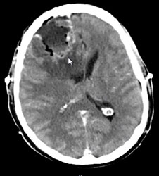
The next image is the immediate postop CT scan. The arrow shows the bottom of the tumor resection bed; above the arrow is some cerebrospinal fluid filling the resection cavity. The brain can be seen to be much less deformed now that the mass is removed.
The tumor was determined by the neuropathologist to be a mixed oligodendroglioma-astrocytoma Grade III. Again, the astrocytic grade is the grade that generally determines the behavior of the tumor, rather than the oligodendroglial portion. The patient received radiotherapy and chemotherapy.
The MRI below shows the patient 7 years later after this treatment with no evidence of any recurrent tumor growth.
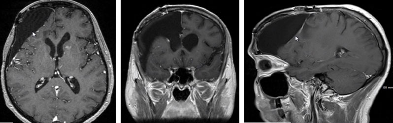
The arrows show the cystic cavity remaining where the tumor was. There is no evidence of recurrence at the time of this MRI 7 years after treatment. The patient continues to function independently and continues to follow up annually with surveillance imaging studies.